New Devices Reveal How Cells React to Stress, Driving Innovations in Biotech and Drug Development

Invented by Pena Castellanos; Brisa, Sbaizero; Orfeo, Abdel-Hafiz; Mostafa, Mestroni; Luisa, Park; Daewon

Cells in our bodies feel different forces every day. These forces, like stretching or squeezing, change how cells work. When this happens in the heart, it can lead to health problems such as scarring or even heart failure. But how do we study these forces in a lab, in a way that really shows what happens inside the body? This is where the cell stressor device comes in. This blog will explain why this device matters, how it stands out from older ideas, and what makes it so unique and helpful in science.
Background and Market Context
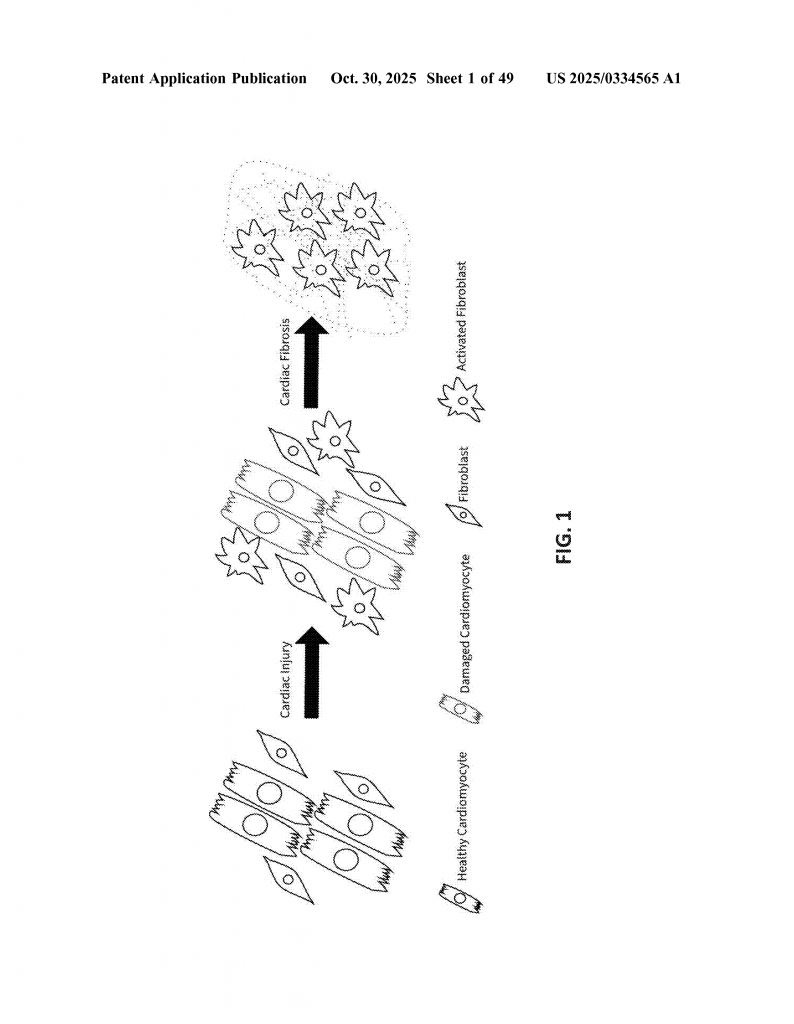
Heart disease is one of the biggest health problems in the world. Every year, millions of people get sick because their hearts do not work well. The heart is always moving, stretching, and squeezing with every beat. When the heart is hurt, like after a heart attack, it tries to fix itself. Sometimes, this leads to scarring inside the heart, which makes it stiff and unable to pump blood well. This scarring is called fibrosis, and it is a big reason why people get heart failure.
Doctors and scientists want to know why this scarring happens and how cells in the heart react to being squeezed or stretched. If they can understand this, they might find new ways to stop the scarring and help people with heart problems. But the problem is that it’s hard to study these forces in a simple and clear way in the lab. Most lab tools either stretch or squeeze cells, but not both at the same time. Also, many old devices are expensive, hard to use, or do not really show what happens in the body.
The cell stressor device was made to solve these problems. It is simple, not expensive, and can be used in regular lab dishes. It can stretch and squeeze cells at the same time, just like what happens in the heart. This makes it very useful for doctors, scientists, and drug companies who want to study heart disease, scarring, or even test new medicines.
Beyond the heart, other organs like the lungs and kidneys also get sick because of scarring from these kinds of forces. If we can use this device to learn more, it could help with many types of diseases, not just heart disease. Since scarring causes about one third of deaths around the world, having a good device for this research is very important.
The market for lab tools that study cells is growing. Scientists want better tools that let them see what really happens in the body. They want tools that are easy to use and do not cost too much. The cell stressor device fits this need very well. Because it can be made by 3D printing and uses simple materials, it is affordable and can be shared with many labs. This opens up research to more people and can speed up finding new treatments for serious diseases.
Scientific Rationale and Prior Art
To understand why this invention matters, let’s look at what came before. Cells feel forces in the body all the time. These forces are called mechanical stresses. There are two main types: tension (stretching) and compression (squeezing). In the heart, both of these happen with every beat. Over time, if the heart is hurt or overworked, these forces can cause the cells to change, leading to scarring and stiffening of the heart wall. Scientists call this remodeling.
For a long time, researchers have tried to copy these forces in the lab. They use devices that stretch rubber sheets or squeeze gels with cells on them. Some devices use special motors or weights. Others use springs, screws, or clamps. But most of these older devices can only do one thing at a time. They might stretch the cells or squeeze them, but not both together. Also, many of these devices are big, hard to use, or cost a lot of money. Some need special parts or cannot be used with regular lab dishes. This makes them less useful for everyday research.
Another problem is that old devices often do not give the same kind of force that happens in real organs. For example, the heart has both tension and compression in close areas at the same time. Old devices might not show this, so the results might not match what happens in people.
There are also technical limits. Some devices use soft rubber or plastic that can change shape over time, so the force is not always the same. Others use materials that are hard for cells to stick to, or they cannot be seen clearly under a microscope. Many require the whole device to be put together before adding the cells, making it hard to do many tests quickly.
Some lab tools use what is called a “four-point bend” test to study materials. This test pushes down on a bar at two points while holding up the ends, making a part in the middle that is stretched and a part that is squeezed. But until now, this idea was not used for studying live cells in a way that is easy, cheap, and works with regular cell culture.
The cell stressor device takes the four-point bend idea and makes it useful for cell research. It uses a small, clear bar where cells can grow. The device can push and pull on this bar, creating both tension and compression at the same time. It can be used in regular lab dishes, uses simple screws or even a small motor, and can be made by 3D printing. This solves many of the problems with old devices.
It is also important to note that the device can be used with many types of cells, including heart cells, lung cells, kidney cells, and even mixed cultures. This means it can help with research in many areas, not just the heart.
Finally, the device is designed so that the amount of force can be carefully measured and repeated. This makes experiments more trustworthy and easier to compare between labs. In short, this invention fills a big gap in the tools available for studying how cells feel and respond to the forces that cause disease.
Invention Description and Key Innovations
The cell stressor device is a simple, clever tool that lets scientists study how living cells respond to being stretched and squeezed. It has a special design based on a “window” made by two ledges and a bar, with a beam and a bender inside. Let’s break down the main parts and what makes it special.
The body of the device is shaped like a tiny frame with two ledges facing each other. The bottom of each ledge sticks to a bar, making a space or window between them. The top of the window is open, so you can slide things in and out. Into this window, you put a thin, flat beam. This beam is made from a material like PMMA, which is smooth, clear, and hard. The top of this beam is where you grow your cells. Before using, the beam is coated with a sticky layer of gelatin or gelatin-fibronectin, so the cells can stick and grow just like in the body.
Under the beam, you place a part called the bender. The bender is smaller than the window, so it fits inside and can move up and down. The bender can be moved by twisting a screw from below, or by using a small motor. When you turn the screw, the bender pushes up on the bottom of the beam. Because the beam sits on the ledges at each end, pushing in the middle makes the top of the beam stretch (tension) and the bottom squeeze (compression) – just like what happens in the heart during each beat.
The device is made so that you can control exactly how much force is used. You can use a special screwdriver that measures torque, or a motor with a sensor, so every experiment is the same. You can even use a computer to control the motor for very fine, repeated movements, letting you mimic the beating of the heart.
Key features include:
– The main body, bender, and beam can be 3D printed, making it easy and cheap to produce.
– The beam is smooth and clear, perfect for growing cells and looking at them under a microscope.
– The device is small enough to fit in a regular petri dish, so it can be used in any biology lab.
– It can be taken apart easily, so you can prepare many beams in advance, or test many samples quickly.
– The shelves inside help hold the beam steady, and allow even force to be applied.
– The design can be changed to make wells for gels or tissue samples, or to add marks for tracking cell movement.
– You can use the device with many types of sensors, like torque or force sensors, to measure exactly what the cells feel.
The device is also easy to sterilize, so it is safe for cell cultures. The beam can be removed after the test and used for staining, imaging, or other analysis. The design allows for live imaging under a microscope, so you can watch how cells move, change shape, or even die when stressed.
The process works like this: you coat the beam, add your cells, and let them grow. Once the cells are ready, you put the beam into the device, add it to a dish with growth medium, and apply the desired force by turning the screw or running the motor. You can then take pictures before and after, or even record video, to see how the cells react. After the experiment, you can use stains to look at the cytoskeleton, see if the cells are alive or dead, measure how stiff they are, and more.
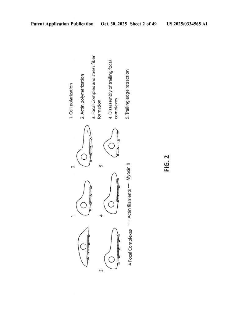
The device has been tested and shown to apply forces in a consistent, measured way. It can apply both stretching and squeezing at the same time, which is what happens in real organs. Tests with heart fibroblast cells show that the cells align, move, or change their internal structure when stressed, just like they do in the body during disease.
The device can be used with many other tools. For example, you can use atomic force microscopy to measure how stiff the cells become after being stressed. You can use confocal microscopy to get clear images of cell structure. You can even use computer programs to track how cells move, or to measure changes in the cell skeleton.
This invention is very flexible. You can make many devices and run many tests at once, making it great for drug testing or research. You can change the size, use different materials, or automate the process. You can even use it to study differences between male and female cells, or compare healthy and sick cells. This makes it a powerful tool for understanding disease and finding new treatments.
In short, the cell stressor device is a breakthrough for studying how cells feel and respond to the forces that cause disease. It is simple, cheap, flexible, and powerful. It brings real-world forces into the lab in a way that is easy to use and easy to trust.
Conclusion
The cell stressor device is a big step forward for research on cell mechanics and disease. It solves many problems found in older tools by making it easy to apply both stretching and squeezing forces to cells at the same time, just like in the body. Its design is simple, affordable, and flexible, making it perfect for labs of all sizes. It can be used to study heart disease, scarring, and many other health problems caused by mechanical forces in cells. By making it easier to understand how cells react to these forces, this device opens the door to better treatments and a deeper understanding of disease. If you are a scientist, doctor, or innovator looking to study cell mechanics, this device is a game changer.
Click here https://ppubs.uspto.gov/pubwebapp/ and search 20250334565.