Enhancing Brain Surgery Precision with Real-Time Neural Mapping and Visual Guidance
Invented by RAPOPORT; Benjamin I., Mermel; Craig H., Hettick; Mark, Poole; Adam J., Reed; Kyle, Forney; Ruth Ann, Ho; Elton, PRECISION NEUROSCIENCE CORPORATION
Treating the human brain is one of medicine’s biggest challenges. Surgeons need to see not just what the brain looks like, but how it works in real time. This new patent application describes a system that helps doctors see both the brain’s structure and its activity during surgery. It uses a special microelectrode array and advanced computer tools to show live brain function over brain images. Let’s break down why this matters, the science behind it, and how the invention works.
Background and Market Context
The human brain is like a busy city. Each part has a different job, and if something goes wrong—like in a stroke, tumor, or injury—figuring out exactly where to operate is tricky. Doctors rely on MRI and CT scans to see the “map” of the brain, but these pictures only show the shape of the brain, not what each part is doing at any moment. This can make brain surgery risky, because if a surgeon touches the wrong spot, it could affect important things like speech, movement, or memory.
For years, doctors have wanted a way to see both the brain’s structure and its live signals at the same time. This would help them avoid damaging important areas and make surgeries safer. Brain-computer interfaces (BCIs) are new tools that can read the brain’s electrical signals. These signals help doctors understand what each part of the brain is doing. Some BCIs use arrays of tiny electrodes placed on the brain’s surface to pick up these signals. The problem is, most tools today either show structure (like an MRI) or function (like electrical signals), but not both together in real time.
The market for these kinds of advanced brain tools is growing fast. As more people need brain surgery for conditions like epilepsy, tumors, or injuries, there’s a strong demand for technologies that make surgery safer and more precise. Hospitals and surgeons want systems that can tell them, moment by moment, what’s happening inside a patient’s brain, so they can make better decisions during operations.
The invention in this patent application aims to solve this need. It brings together a high-density microelectrode array and a computer system that overlays brain signals onto brain images in real time. This means that during surgery, doctors can see both the map of the brain and the live activity overlaid together, like a weather map showing storms moving across a city.
Scientific Rationale and Prior Art
To understand why this invention is important, let’s look at the science behind brain mapping and what has been tried before.
The brain works by sending tiny electrical signals between cells called neurons. Scientists can measure these signals with electrodes. There are two main ways to do this: one is by poking tiny electrodes into the brain; the other is by placing them on the surface. Poking into the brain can give very detailed information, but it can also damage the brain and is hard to do safely, especially in humans. Surface electrodes are gentler and safer, but older versions did not pick up signals as clearly or with as much detail.
Recently, advances in materials science have made it possible to create arrays with hundreds or even thousands of tiny electrodes on a flexible sheet. These high-density microelectrode arrays can sit on the brain’s surface and pick up detailed signals without poking into the tissue. These signals are called electrocorticography (ECoG) signals. When the electrodes are really close together and small, we call it micro-ECoG (μECoG). Scientists have shown that μECoG can pick up detailed patterns of brain activity, which are useful for guiding surgery or for brain-computer interfaces.
But there’s a big gap: even though we can record these signals, most surgical navigation tools still only show static images from MRIs or CTs. They don’t let surgeons “see” the live brain activity mapped onto those images in real time. Functional MRI (fMRI) can show which brain areas are active, but it is slow and not very detailed. It also can’t be used during surgery.
Some systems have tried to combine functional and structural information, but they are either too slow, not detailed enough, or not practical for use during an operation. Plus, getting the position of the electrode array exactly right on the brain (so you know what part of the image matches what you’re recording) is hard. Existing systems might use simple markers, but they often don’t track the array’s position with enough accuracy or update the display in real time as the brain shifts during surgery.
In summary, the main problems with earlier approaches are:
– Slow or low-detail imaging of brain function (like fMRI).
– Lack of real-time updates during surgery.
– Hard to accurately match recorded signals to brain images.
– Electrode arrays that are either too invasive or not detailed enough.
– No easy way for surgeons to see both brain structure and function together, live, during operations.
This patent builds on recent breakthroughs in flexible, high-density electrode arrays and computer vision. It uses special markers (fiducials) on the array and advanced tracking (optical or electromagnetic) to know exactly where the array is at all times. The computer system can then overlay live brain signals onto the brain image, updating in real time as the procedure goes on. This is a leap forward from anything that has come before.
Invention Description and Key Innovations
Now, let’s look at how this new system works and what makes it so special.
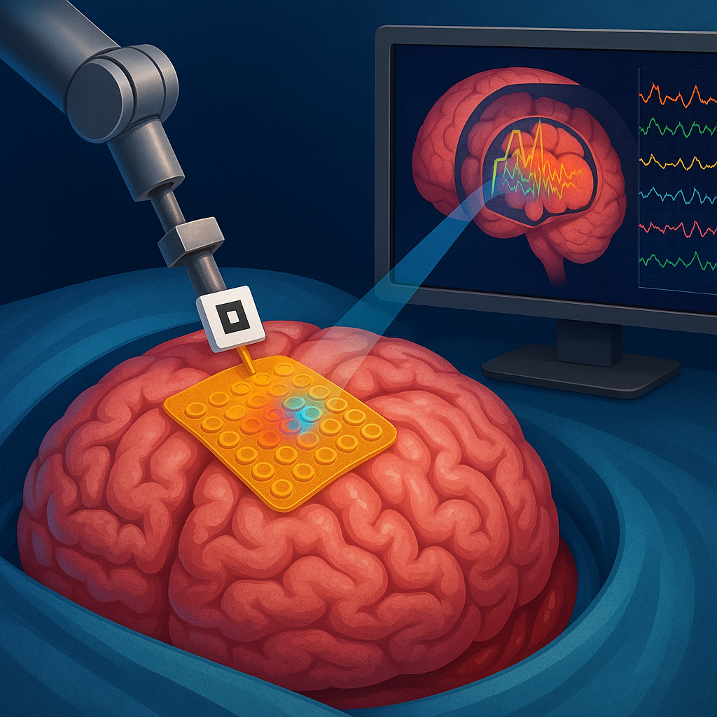
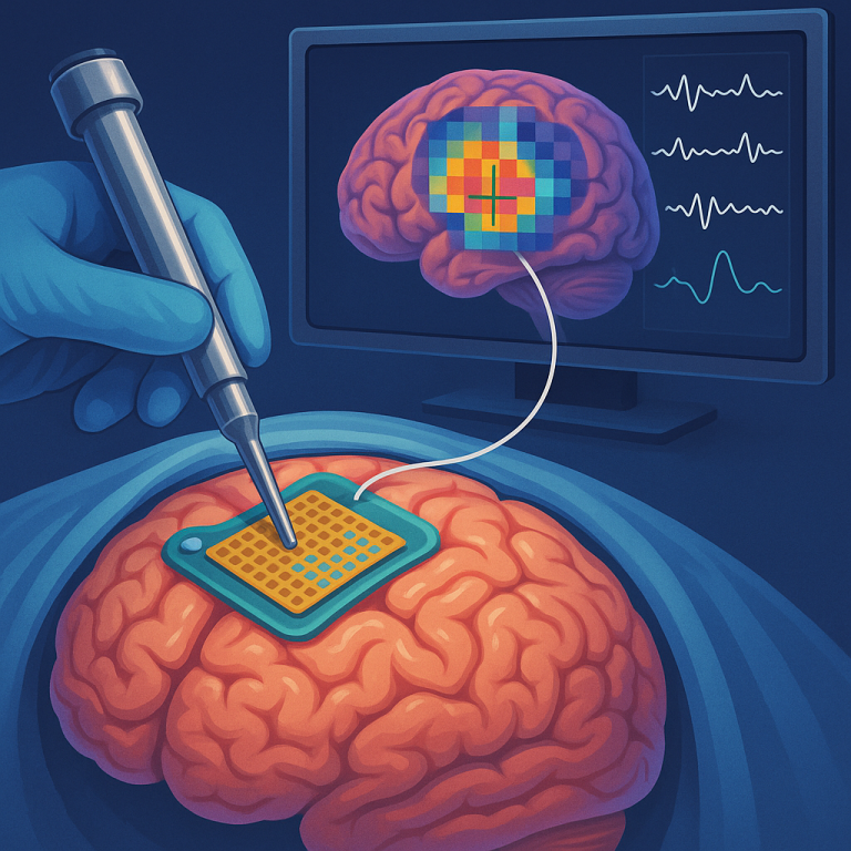
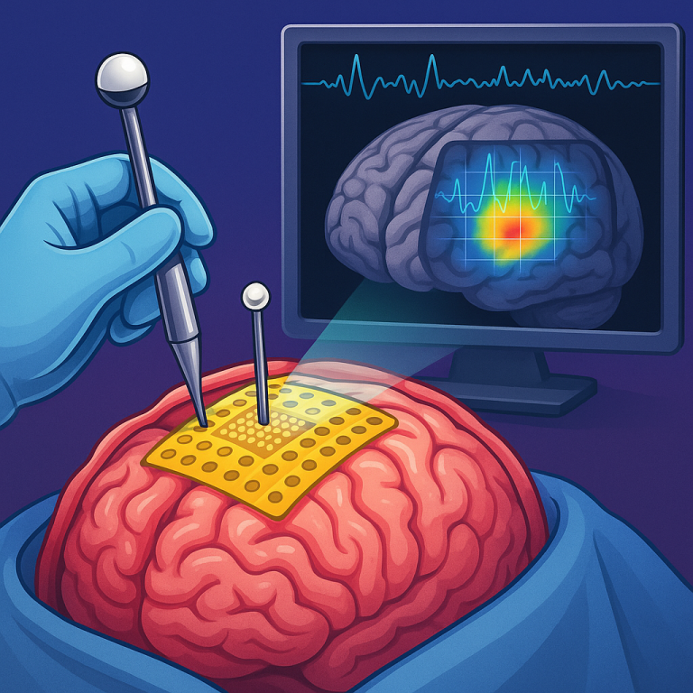
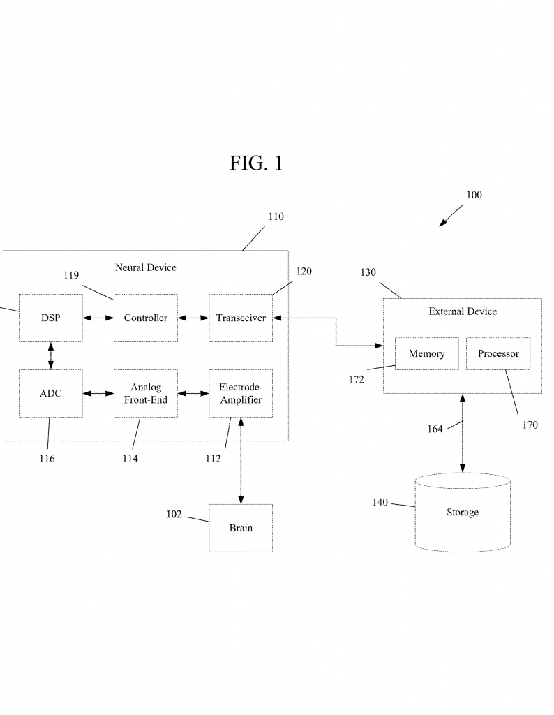
At the heart of the invention is a microelectrode array—a very thin, flexible sheet covered with thousands of tiny electrodes. This array is designed to be placed gently on the surface of the brain during surgery. Unlike older arrays, it does not poke into the brain, so it is safer and causes less harm. The sheet can bend and fit the shape of the brain, which means it maintains good contact and picks up strong, clear signals from many areas at once.
What sets this array apart is the use of special markers, called fiducials. These are like little “checkpoints” that the computer system can “see” using cameras or other sensors in the operating room. The fiducials might be special shapes, codes, or materials that show up clearly in images or scans. They let the system track the exact position and orientation of the array on the brain. This is important, because brains can shift during surgery, and it’s critical to know exactly where each electrode is.
The system includes a powerful computer with a special software program. The computer’s job is to:
– Register, or “lock in,” the array’s position on the brain, using the fiducials and tracking systems (optical or electromagnetic).
– Receive real-time electrical signals from each of the thousands of electrodes.
– Process these signals to find patterns, like areas of high activity or certain rhythms.

– Overlay this functional data onto images of the patient’s brain (from MRI or CT scans).
– Display this combined view to the surgeon in real time, updating instantly as new signals come in or as the array moves.
The visualization can take forms like heatmaps (where colors show activity levels), dot plots, or even time-frequency spectrograms (which show how the brain’s rhythms change over time). The surgeon can see exactly which part of the brain is most active, and how that matches up with the anatomy. If the array is moved, or if the brain shifts, the system updates automatically.
The surgeon also gets tools to select specific electrodes or groups of electrodes, look closer at certain brain regions, and even record and play back the signals for later review. The system can process signals to find “oscillatory patterns”—these are the brain’s natural rhythms, which can tell the surgeon if a part of the brain is responsible for movement, speech, or something else.
One of the most important features is real-time updating. If something changes during surgery—say, the surgeon moves the array, or the brain shifts a bit—the computer notices right away and updates the display. This way, the information is always current, helping the surgeon make the best decisions and avoid critical areas.
The system can also be used for many types of brain surgery. For example, when removing a tumor, the surgeon needs to know where language or movement centers are so they can avoid them. The system helps find those areas by showing live activity. It can also help with implanting brain-computer interfaces for paralyzed patients, or with mapping areas responsible for seizures in epilepsy surgery.
The hardware is designed to be safe, minimally invasive, and easy to use. The microelectrode array can have over 1,000 electrodes, each only a fraction of a millimeter wide. The flexible material means it can cover curved parts of the brain without hurting the tissue. The computer system is built to process and visualize huge amounts of data quickly, so there’s no lag or delay during surgery.
The invention also includes special software tools for checking signal quality, measuring impedance, and identifying noise or interference. This helps make sure the data shown to the surgeon is accurate and reliable. The user interface is designed so that both surgeons and their technical helpers can easily interact with the system, adjust settings, and annotate important findings. Everything is built with the needs of the operating room in mind: speed, clarity, and safety.
Another important innovation is the use of tracking technology. By using optical or electromagnetic trackers, the system can “see” not just the array, but also surgical tools, the patient’s head, and other items in the room. This allows for very precise mapping of everything involved, helping keep the display aligned with reality even if things move.
The system can also use machine learning to help process the neural signals. As more data is gathered, it can learn to spot important patterns automatically, or even alert the surgeon to changes that might be important. This makes the system even smarter and more helpful over time.
To sum up, the key innovations in this invention are:
– A thin, flexible, high-density microelectrode array that can record over 1,000 channels at once.
– Built-in fiducials for accurate, real-time tracking of the array’s position on the brain.
– A computer system that receives, processes, and overlays live neural signals onto brain images, updating in real time.
– Tools for signal quality checking, visualization, and selection of specific electrodes or regions.
– Integration with optical or electromagnetic tracking for precise mapping, even as things move during surgery.
– Machine learning features for smarter data processing and alerting.
This system brings together the best in brain mapping, imaging, and user-centered design to give surgeons a clear, always current picture of both the brain’s anatomy and activity. It stands to make brain surgery safer, more precise, and more effective.
Conclusion
This new neuronavigation system represents a big step forward in brain surgery. By letting surgeons see both the map and the live activity of the brain at once, it gives them the information they need to operate with more confidence and care. The combination of a flexible, high-density electrode array, smart tracking, and real-time visualization makes this tool a valuable addition to the field. It can be used in many types of neurosurgery, from removing tumors to implanting brain-computer interfaces, and stands to improve outcomes for patients everywhere. As hospitals and clinics look for better ways to treat brain conditions, systems like this will be at the forefront of safe, smart, and personalized care.
Click here https://ppubs.uspto.gov/pubwebapp/ and search 20250331928.