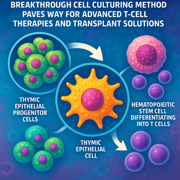
Breakthrough Cell Culturing Method Paves Way for Advanced T-Cell Therapies and Transplant Solutions
Invented by HAMAZAKI; Yoko, PRETEMER; Yann, GAO; Yuxian, Kyoto University
This article will help you learn about a new way to make thymic epithelial cells, which are important for the immune system. We will clearly explain the background, the science behind this invention, and describe the new method in simple words. By the end, you will understand why this breakthrough matters for medicine and health.
Background and Market Context
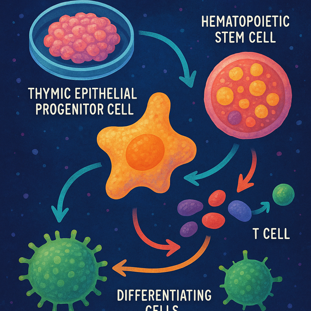
The immune system protects our bodies from germs, cancers, and other threats. T cells are a key part of this system. They learn to identify and attack harmful things inside a special organ called the thymus. The thymus is like a school for T cells. Inside, special helper cells called thymic epithelial cells (TECs) teach T cells what to fight and what to ignore. As people get older, or after some medical treatments, the thymus stops working as well. This makes the body weaker against sickness and affects recovery after some treatments.
Many people around the world face problems because their immune system does not work well. This can happen because of age, disease, or after treatments like chemotherapy. When the thymus is damaged or stops working, people are more likely to get infections, develop autoimmune diseases, or have trouble fighting off cancer. Scientists and doctors want to find ways to help these people by repairing or replacing parts of the immune system.
One idea is to grow new TECs in the lab. If doctors can make TECs from a patient’s own cells, they could help “reboot” the immune system. This could be useful for older people, for children born with immune problems, and for patients whose immune system has been harmed by treatment. However, making mature, working TECs in a dish has been very hard. So far, scientists could make early versions of TECs, called progenitors, but they could not make fully developed TECs outside the body. This has been a big problem for turning the idea into real treatments.
If someone finds a way to make mature TECs in the lab, it could open up new treatments for many diseases. It could help people recover after bone marrow transplants, fight off cancers, avoid harmful immune attacks, and recover from infections. There is a large market for such therapies, and big interest from hospitals, biotech companies, and patients. The demand will likely grow as more people live longer and as more treatments can harm the immune system.
Scientific Rationale and Prior Art
To understand the new method, we need to look at how TECs develop both in the body and in the lab. In the body, TECs come from early cells called stem cells, which first turn into a layer called the endoderm. The endoderm then forms the foregut, a tube that later makes organs like the lungs and thymus. Some cells in the foregut become pharyngeal endoderm cells, and from there, some of these turn into thymic epithelial progenitors (TEPs). These TEPs can become mature TECs. The process is guided by signals from the body, including special molecules like retinoic acid and FGF-8.
Earlier research could make TEPs from stem cells in the lab by carefully adding certain factors. These include retinoic acid, which helps form the right part of the endoderm, and FGF-8, which tells cells to grow and change. After making TEPs, scientists tried to make them become mature TECs. However, they usually had to put the cells into a living animal (like a mouse) to finish the process. In other words, it was not possible to get fully working TECs just by using dishes and chemicals outside the body.
Several scientific papers and patents have described ways to get TEPs from human or mouse stem cells. These methods often use three-dimensional (3D) cultures or organoids, which are mini-organs grown in the lab. The TEPs made this way showed some signs of being TECs, but they were not fully mature and could not do all the jobs real TECs do. Some papers reported that to get mature TECs, the TEPs had to be transplanted into an animal, where they would finish maturing in a living environment.
The main reason for this problem is that the last steps of TEC development seem to need signals or factors that are hard to copy in a simple dish. Scientists thought that adding more growth factors, or making the culture more like a real organ, would help. But in practice, these tricks did not work well. No one had shown that you could get mature, working TECs just by letting TEPs grow in a dish for a long time with simple conditions. This was the missing step needed for practical therapies.
The new invention tries a different approach. Instead of adding more factors, it removes them. The inventors found that if you stop giving the cells certain factors (like retinoic acid and FGF-8) after a certain point, and just let the TEPs grow for a long time, they slowly mature into real TECs. This is surprising, as most previous work focused on adding more signals, not taking them away. The inventors also found that by sorting cells based on a marker called FOXN1, they could pick out the most mature TECs. This new method is different from past work, which relied on animal grafts or complex mixtures of factors.
Invention Description and Key Innovations
The invention provides a new way to make mature thymic epithelial cells (TECs) from stem cells, all outside the body. The process has several steps, each designed to match how cells would develop in a real thymus, but using simple lab conditions.
First, the inventors start with pluripotent stem cells. These are cells that can turn into almost any type of cell. They coax these stem cells to become anterior foregut endoderm cells by using certain mixtures in the dish. This is done by adding specific chemicals (like Activin A and others) for a set number of days, following known recipes.
After this, the next step is to turn these foregut cells into pharyngeal endoderm cells. This is done by adding retinoic acid (RA) to the culture. Sometimes, FGF-8 is added as well, to help guide the cells. The process is carefully timed and the amount of RA is controlled to get the right kind of endoderm, which is needed for thymus formation.
When the pharyngeal endoderm cells are ready, the next step is to make TEPs. Here, the inventors do something new: instead of adding more growth factors, they remove them. They stop giving the cells RA and FGF-8, and continue to culture them in a simple medium. Over the next several days, the cells start to turn into TEPs, which are early TECs. These TEPs are marked by the appearance of a gene called FOXN1, which is a key signal that the cells are on the right path.
The final and most important step is to let the TEPs grow for a long time—at least 25 days, often much longer, up to 80 or even 110 days. During this time, no added RA or FGF-8 is provided. In fact, the inventors say that the process works best when other signals (like sonic hedgehog, BMP-4, and noggin) are also left out. The cells are just given a basic medium that keeps them alive but does not push them in any direction.
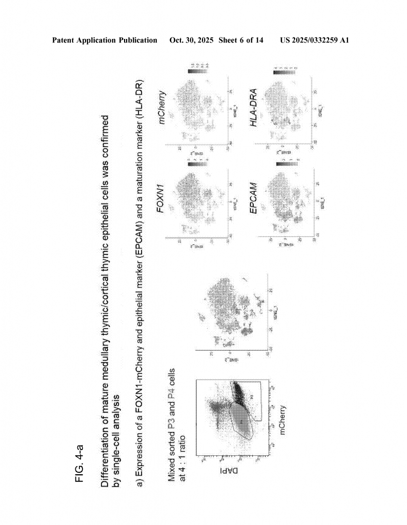
As the days go by, the TEPs slowly change into mature TECs. This happens naturally over time, without extra signals. The inventors found that the cells begin to express more of the markers that real TECs have, like HLA-DRA and others important for immune function. They also found that by sorting for cells that have high levels of FOXN1, they could pick out the most mature TECs, which have the best ability to help T cells develop.
The new method can be done without feeder cells (other cells that help in culture), and without any animal products, making it safer and better for future medical use. This is called “feeder-free” and “xeno-free” culture. This is important for making treatments that can be given to humans.
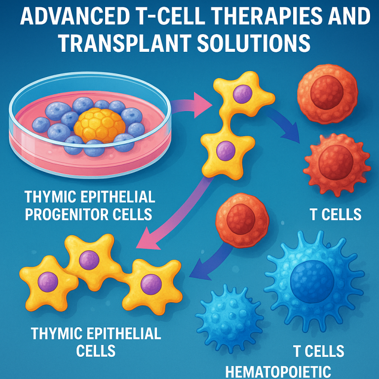
Once the mature TECs are ready, they can be used in several ways. They can be transplanted into patients to repair or replace the thymus. They can also be mixed with blood stem cells to help make new T cells in the lab. This is done by putting the TECs and blood stem cells together in a dish and letting them interact. The result is the production of new, functional T cells, including all the major types needed for health (CD4, CD8, and others).
The inventors showed that the T cells made this way have a wide variety, just like real T cells in a healthy person. This is important, because it means the method can make a full, working immune system—not just a few types of T cells. The diversity of T cell receptors (TCRs) was checked and found to be similar to that in normal blood.
There are other technical details that make this invention stand out. The inventors use careful timing, defined media, and genetic markers to follow the cells’ progress. They establish special reporter cell lines (using a red fluorescent protein called mCherry linked to FOXN1) to track which cells are becoming TECs. They use flow cytometry and gene expression tests to sort and verify the maturity of the TECs they make.
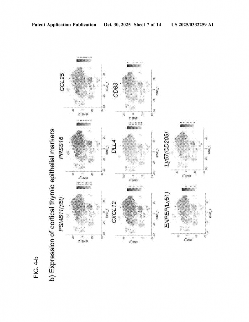
This method also allows the creation of both main types of TECs: cortical and medullary. These two types are needed for all the steps of T cell education. Most past methods could only make immature or partial TECs. Here, the inventors show that both types can be made and identified using markers.
The new method is flexible and can be adapted for use with human or animal cells. It can be done with different kinds of stem cells, including iPS cells (from adult tissues) and ES cells (from embryos). This means it has the potential for wide use in research, therapy, and even drug testing.
Lastly, the invention provides not just a method, but also the actual cells produced by the method, transplantation materials made from these cells, and T cells made using the TECs. It covers the full chain from stem cells to immune therapy, making it a broad and practical solution for immune system repair.
Conclusion
This new method for making thymic epithelial cells is a big step forward. It solves a long-standing problem by allowing mature TECs to be made outside the body, using simple and safe lab methods. This breakthrough could help people with weak immune systems, support better recovery after cancer treatment, and open new doors for treating autoimmune diseases. By using the body’s own cells and guiding them with careful steps, the method avoids the need for animal testing or complicated mixtures of chemicals. It is a clean, reliable, and practical solution.
The power of this invention is in its simplicity and the way it follows the natural path of cell development, but with the clever twist of removing signals rather than adding more. This shows that sometimes, letting nature take its course—with just the right starting push—leads to the best results. As research continues, we can expect to see new therapies based on this method, helping patients of all ages regain a healthy immune system.
Click here https://ppubs.uspto.gov/pubwebapp/ and search 20250332259.